Figure 6.
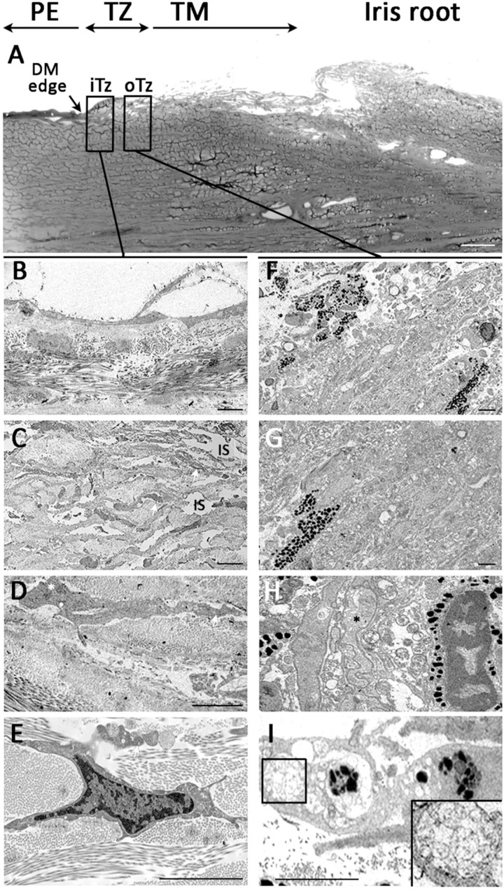
Ultrastructural morphology under transmission electron microscopy. (A) Overview picture illustrates posterior limbus with peripheral endothelium (PE) with DM edge, transition zone with inner and outer TZ, followed by trabecular meshwork (TM) and iris root. Inner TZ. (B,C) Cells in the loosely arranged stromal matrix with numerous interstitial spaces (IS). (D,E) The cells had high nuclear/cytoplasmic ratio and with loose chromatin and pronounced nucleoli (white asterisk in C). Outer TZ. (F,G) A mix of non-pigmented and pigmented cells in a closely packed matrix. (H) Cells containing cytoplasmic granules with pigmentation adjacent to highly convoluted non-pigmented cells. (I) Cell with phagosomes containing irregular deposits (magnified image in inset). Abbreviations: iTZ: inner TZ; oTZ: outer TZ; DM: Descemet’s membrane. Scale bars: 50 μm (A); 10 μm (B–F, I); 5 μm (G).

