Figure 4.
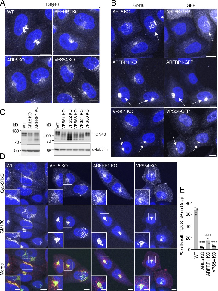
Altered localization of TGN46 and impaired transport of STxB to the Golgi complex in ARL5-KO and ARFRP1-KO cells. (A) Immunofluorescence microscopy of WT, ARFRP1-KO, ARL5-KO, and VPS54-KO cells immunostained for endogenous TGN46 and counterstained with DAPI (blue). Scale bars: 10 μm. (B) ARFRP1-KO, ARL5-KO, or VPS54-KO cells were rescued by expression of the corresponding GFP-tagged proteins, stained for endogenous TGN46, transgenic GFP (only for VPS54-GFP), and nuclei (DAPI; blue) and imaged by confocal microscopy (GFP fluorescence was directly observed for ARL5B-GFP and ARFRP1-GFP). Scale bars: 10 μm. Arrows indicate rescued cells. (C) SDS-PAGE and immunoblot analysis of endogenous TGN46 and α-tubulin (loading control) in WT and the indicated KO cells. The positions of molecular mass markers are indicated on the left. (D) Live WT, ARL5-KO, ARFRP1-KO, or VPS54-KO cells were incubated for 15 min with Cy3-STxB and chased for 1 h in regular culture medium at 37°C, after which cells were fixed, immunostained for endogenous GM130, and imaged by confocal microscopy. Scale bars: 10 μm. Insets are magnified views of the boxed areas. Inset scale bars: 5 μm. (E) Quantification of cells having Cy3-STxB staining at the TGN as described in Fig. 2 C. Values are the mean ± SEM from three independent experiments. More than 100 cells per sample were counted in each experiment. ***, P < 0.001, in comparison to WT cells using Dunnett’s test.

