Figure 5.
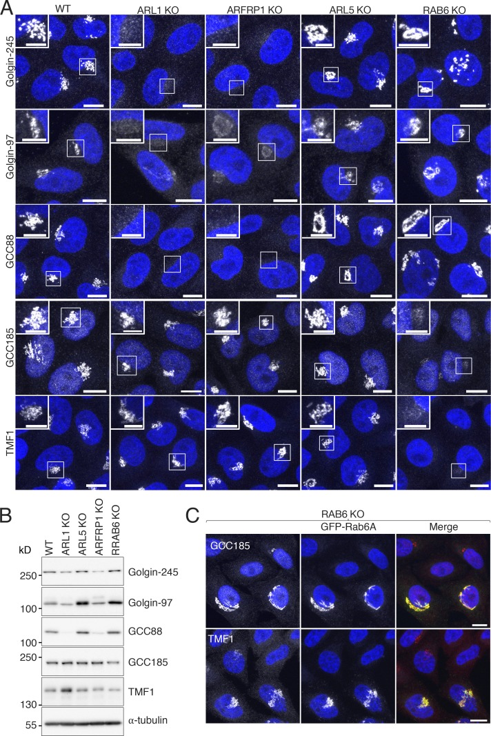
Small GTPases required for localization of golgins to the TGN. (A) Immunofluorescence microscopy of WT, ARL1-KO, ARFRP1-KO, ARL5-KO, and RAB6-KO cells immunostained for endogenous Golgin-245, Golgin-97, GCC88, GCC185, or TMF1 and counterstained with DAPI (blue). Scale bars: 10 μm. Insets are magnified views of the boxed areas. Inset scale bars: 5 μm. Notice that ARL1 KO or ARFRP1 KO caused complete disappearance of Golgin-245 and GCC88, and a partial decrease in the intensity of Golgin-97, at the TGN; quantification in 10 cells per sample in three independent experiment showed that Golgin-97 decrease was 75.6% ± 2.4% in ARL1-KO cells and 55.0% ± 1.9% in ARFRP1-KO cells. (B) SDS-PAGE and immunoblot analysis of endogenous golgins and α-tubulin (loading control) in WT and KO cells. The positions of molecular mass markers are indicated on the left. (C) Immunofluorescence microscopy of RAB6-KO cells transfected with a plasmid encoding GFP-tagged mouse Rab6A (green), immunostained for endogenous GCC185 and TMF1 (red), and counterstained with DAPI (blue). Cells were examined for GFP fluorescence by confocal microscopy. Scale bars: 10 μm.

