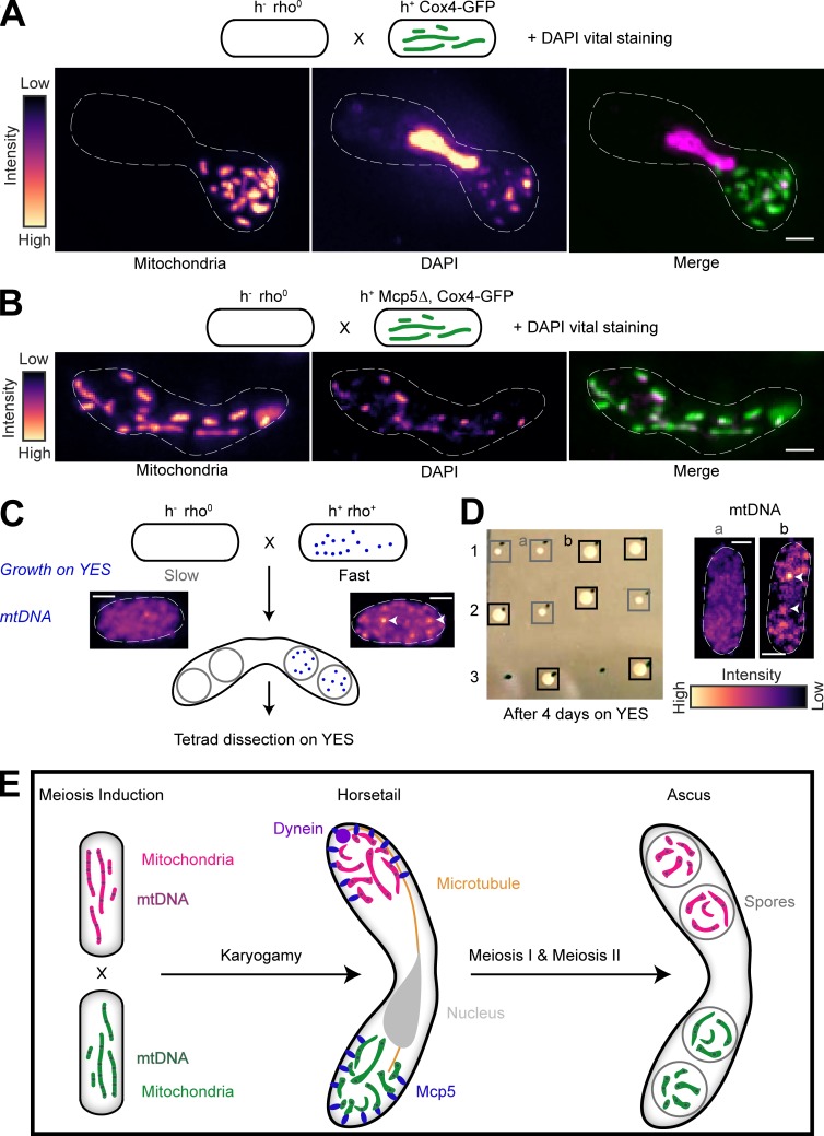
Figure 6.
mtDNA are uniparentally inherited during fission yeast meiosis. (A) Schematic of the cross and DAPI vital staining performed (top, strain PHP14xPT1650; see Table S1), maximum-intensity–projected images of mitochondria labeled with Cox4-GFP (left) and mtDNA (DAPI, center) represented in the intensity map to the left of the images and their merge (right). Note that a small portion of zygotes exhibit nuclear DAPI signal during vital staining. One such zygote has been chosen here to demonstrate that the mitochondria and mtDNA remain segregated even upon complete fusion of the zygote, as indicated by the horsetail nucleus. (B) Schematic of the cross and DAPI vital staining performed (top, strain PHP14xVA074; see Table S1), maximum-intensity–projected images of mitochondria labeled with Cox4-GFP (left) and mtDNA (DAPI, center) represented in the intensity map to the left of the images, and their merge (right). (C) Schematic of the rho0 and rho+ cross performed to obtain asci, followed by tetrad dissection and growth on YES plates. Maximum-intensity–projected images of vital DAPI staining show absence of mtDNA in rho0 cells (left) and presence in rho+ cells (right, white arrowheads). Note that rho0 cells grow slower than rho+ cells on rich medium. (D) Image of colonies formed on YES (left) following tetrad dissection of asci formed from the cross indicated in A, and maximum-intensity–projected images of vital DAPI staining (right) in representative slow-growing (gray boxes, a) and fast-growing (black boxes, b) colonies, with mtDNA indicated with white arrowheads. (E) Schematic of uniparental mitochondrial inheritance in fission yeast mediated by the tethering of parental mitochondria to the cortex by the anchor protein Mcp5. In A–D, scale bars represent 2 µm and dashed lines represent cell outlines.

