Figure 6.
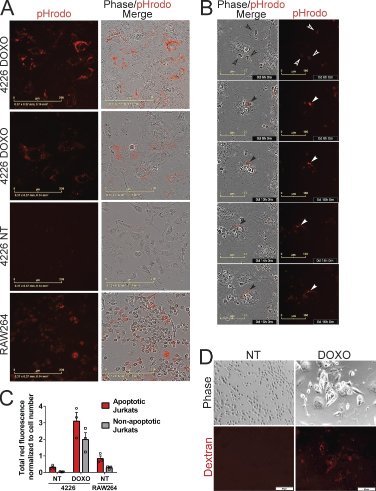
Doxorubicin (DOXO)-induced senescent cells are proficient in phagocytosis. (A–C) pHrodo-labeled Jurkat cells that were NT (nonapoptotic) or treated with doxorubicin for 24 h (apoptotic) were placed onto cultures of RAW264 macrophages and proliferating and senescent 4226 cells. After 2 d, Jurkats were washed off and plates were imaged and quantitated on IncuCyte for the red fluorescence emitted after the phagocytosis of Jurkat cells and their transport to lysosome. (A) Representative images of doxorubicin-treated, senescent 4226 cells (DOXO), NT 4226 cells, and RAW264 macrophage cell line. Scale bar represents 200 µm. (B) Representative phase and red fluorescence images were captured over a time course of a senescent 4226 cell engulfing pHrodo-labeled cells and processing them to the lysosome. Scale bar represents 100 µm; time of capture after addition of pHrodo Jurkats is shown. Open arrowheads indicate pHrodo Jurkats before engulfment, and closed arrowheads indicate pHrodo fluorescence activation at low pH, such as occurs in the lysosome. (C) Total red fluorescence in each well was quantified and normalized to the cell number in each well. The mean and individual data points from triplicate wells were plotted; error bar represents SEM. (D) Proliferating and senescent 4226 cells were serum starved for 1 h, 60 µg/ml of 70-kD Texas Red Dextran was added, 30 min later cells were washed and fixed with 4% formaldehyde, and images were captured. Representative images shown. Scale bar represents 20 µm. Data shown are representative of at least two independent experiments.

