Figure 1.
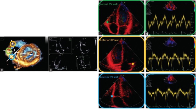
Transthoracic three-dimensional echocardiography, documenting the en-face short-axis view to the mitral valve from the LV cavity, is used only for demonstrative purposes. In white, the cutting planes of RV are labeled. The green circle indicates the lateral RV wall, the yellow circle the anterior RV wall, and the blue circle the inferior RV wall (a). Triplane imaging of the RV was obtained, focusing on RV using the apical four-chamber view as the primary scan plane (b). Complementary views of RV using TDI in addition to the conventional standardized RV views were recorded (c). The sample volume of pulsed-wave TDI was placed on the RV lateral, anterior, and inferior wall at the lateral, anterior, and inferior tricuspid annulus (in green, yellow, and blue, respectively). Tissue velocities were obtained from the three RV walls, respectively (d). LV = Left ventricular, RV = Right ventricle, TDI = Tissue Doppler imaging

