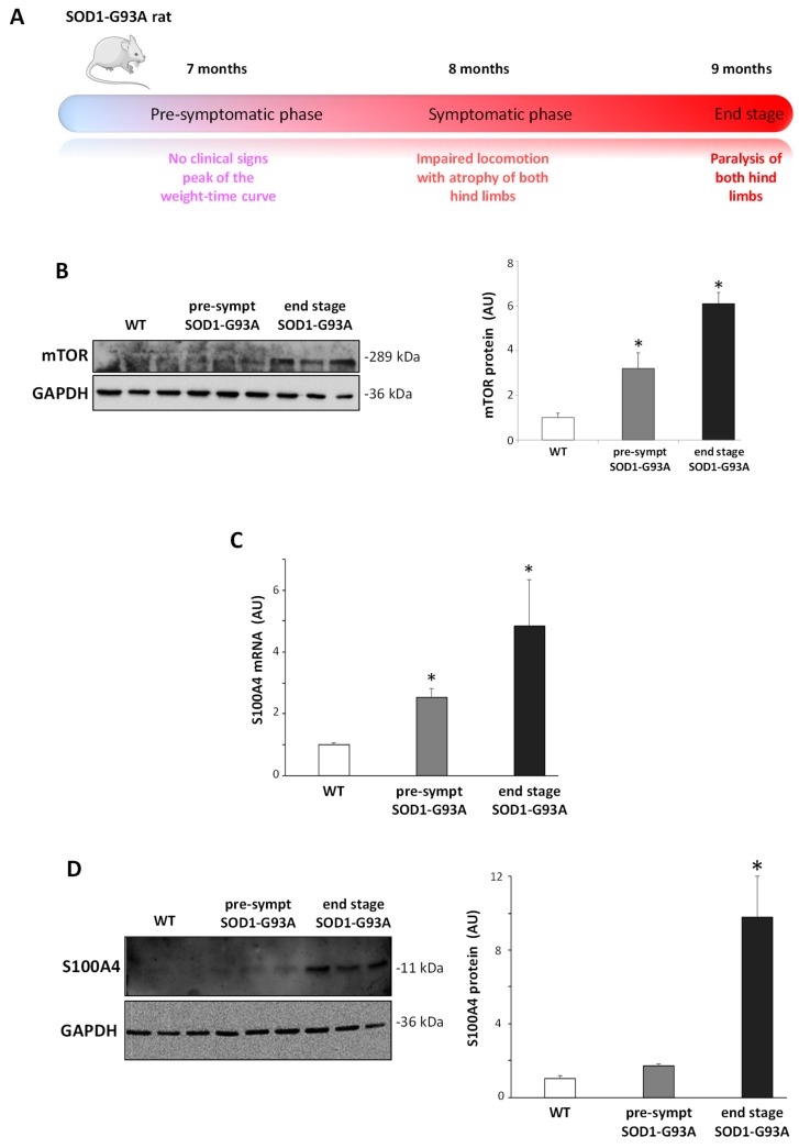
Figure 5.
Increase in the S100A4 mRNA and protein in the lumbar spinal cord from pre-symptomatic and end-stage SOD1-G93A rats. (A) Schematic illustration of the disease development and progression in the SOD1-G93A transgenic rats. (B) Protein lysates of the lumbar spinal cord from wild-type (WT), pre-symptomatic, and end-stage SOD1-G93A rats were analyzed by western blot with the anti-mTOR antibody. (C) mRNA from the lumbar spinal cord from WT, pre-symptomatic, and end-stage SOD1-G93A rats was analyzed by RT-qPCR, and quantification expressed in arbitrary units (AU), relative to WT animals. (D) Protein lysates of the lumbar spinal cord from WT, pre-symptomatic, and end-stage SOD1-G93A rats were analyzed by western blot with the anti-S100A4 antibody. Anti-GAPDH antibody was used to normalize the samples. The graph shows signal quantification expressed in arbitrary units (AU), relative to WT animals. Data are reported as mean ± S.E.M. (n = 3 animals per group). Statistical significance was calculated by ANOVA followed by Post Hoc Tukey’s test. * p < 0.05 relative to WT group.

