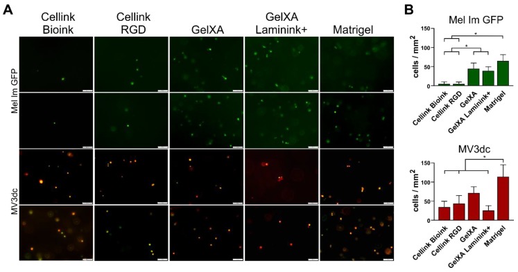
Figure 2.
Survival of melanoma cells in the bioinks. (A) Two representative fluorescence microscope images of each of the cell lines Mel Im GFP and MV3dc one day after 3D printing. Both melanoma cell lines survived the bioprinting and crosslinking process in all bioinks. Scale bars represent 100 µm. (B) Quantification of living cells per mm2 in the bioinks on the day Mel Im GFP showed low amounts of living cells in both Cellink-based inks, a higher rate in GelXA-based inks, and revealed the significantly highest amount of viable cells in Matrigel. MV3dc revealed appropriate amounts of living cells in all materials, with the lowest amount in the Cellinks and GelXA Laminink+, and the significantly highest amount in Matrigel. * p ≤ 0.05 (One-way ANOVA).

