Figure 3.
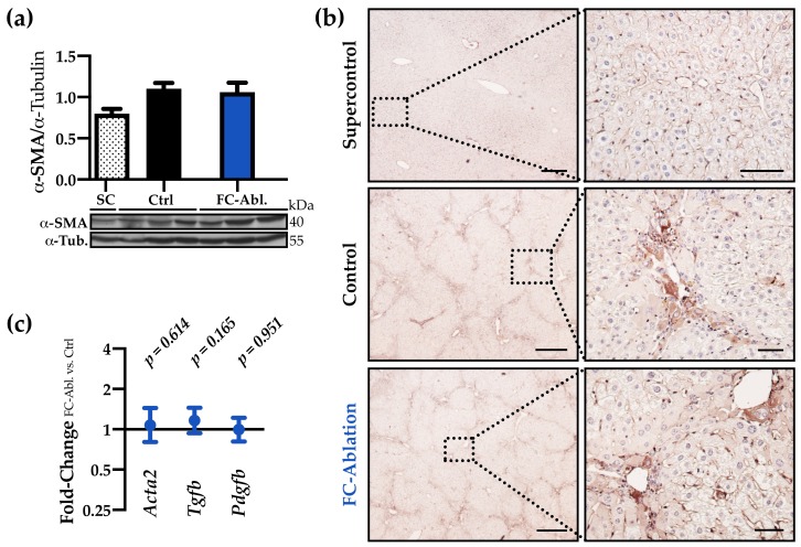
The antifibrotic effect was not accompanied by a reduction of myofibroblasts. (a) Western blot analysis and optical densitometry thereof revealed that the hepatic α-SMA levels were increased following TAA-treatment but unchanged by fibrocyte ablation. Two individual western blots were included in the analysis, a representative blot is shown. Arbitrary unit. SC n = 2; Ctrl, FC-Abl. n = 6. Mean + SEM is depicted. (b) Immunohistochemical staining of α-SMA (brown) demonstrated the periportal accumulation of myofibroblasts in TAA-treated animals and an unchanged expression pattern in result of the fibrocyte ablation. Representative stainings are shown. Magnification 40× and 200×, bars 400 and 50 µm. (c) Hepatic gene expression levels of Acta2, Tgfb, and Pdgfb were comparable at the end of the experiment (full data in Figure S5a–c).

