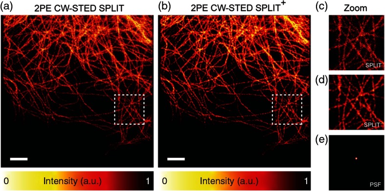
Fig. 4.
(a), (b) 2PE CW-STED SPLIT imaging of -tubulin cells labeled with Alexa 488. The quality of the images is further enhanced applying deconvolution (Richardson–Lucy) on the SPLIT image. (c)–(e) The magnified views of the marked area. (e) The PSF imaging used on the deconvolution. Scale bars: .

