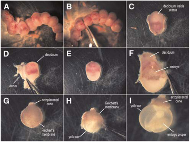
FIGURE 1.

Dissection of an E9.5 mouse embryo. The uterine horn is removed from the mother and separated into individual embryos surrounded by the uterus and deciduum (A–C). The uterus is peeled away (D), leaving the embryo surrounded by a thick decidual layer (E). The deciduum is then removed (F) and the Reichert’s membrane/trophoblast layer (G,H) is carefully separated from the embryo, exposing the intact embryo with the yolk sac available for live imaging (I).
