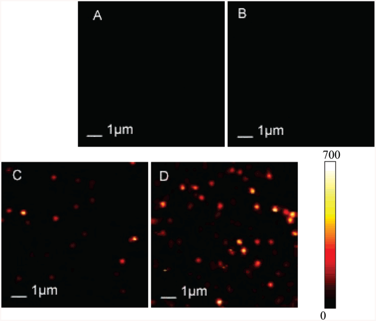
Figure 2.
Typical 10 × 10 μm fluorescence images. These two-dimensional images are 150 × 150 pixels and were acquired in less than 1 min. Each pixel has a 0.6-ms dwell time, and the fluorescence intensity is displayed in a colorized scale, ranging from dark to light. (A) Cy5-labeled dsDNA molecules spin-dispersed on a glass coverslip; (B) silver island film deposited on a glass coverslip; (C) Cy5-labeled dsDNA-SH molecules tethered to silver island film deposited on a glass coverslip with an incubation concentration of 0.5 nM; (D) Cy5-labeled dsDNA-SH molecules tethered to silver island film deposited on a glass coverslip with incubation concentration of 1.5 nM. Illumination intensity was maintained at 1 μW.

