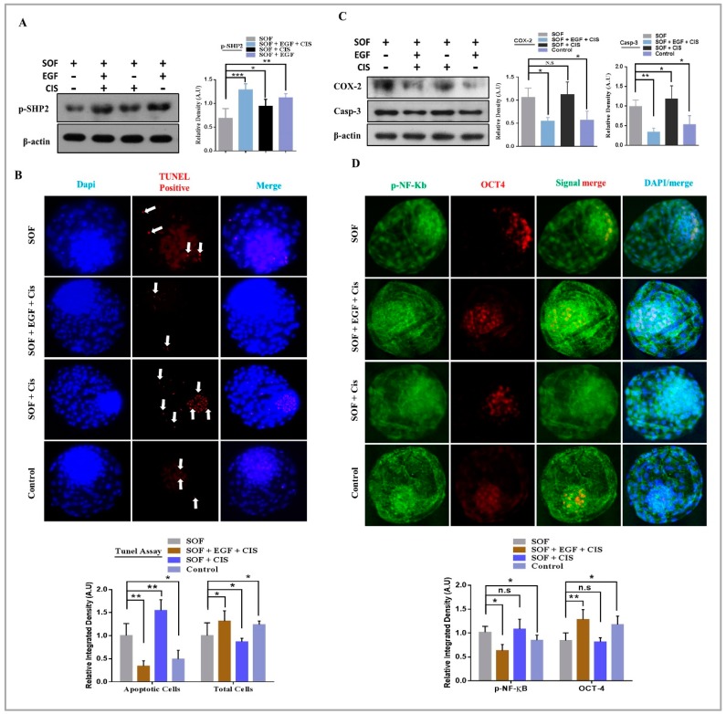
Figure 3.
EGF neutralize cisplatin induced apoptosis and enhances the developmental rate of blastocysts. (A) Western blot analysis of SHP2 in blastocysts grown in SOF, COMBO, CISPLATIN and Vehicle-treated CONTROL media (n = 20 per each group). β-actin was used as a loading control for western blot. The bands were quantified using ImageJ software, and the differences are represented by histogram. (B) TUNEL assay was performed to detect apoptotic cells. TUNEL positive cells were markedly enhanced in CISPLATIN group and were mostly present in the ICM as compare to SOF, COMBO and vehicle-treated CONTROL groups (n = 20 per each group). (C) Western blot of COX-2 and Caspase-3 protein expression showing high apoptosis in CISPLATIN group as compare to other groups (n = 20 per each group). β-actin was used as a loading control for western blot. The bands were quantified using ImageJ software, and the differences are represented by histograms. (D) Blastocysts were costained with OCT4 (red) and p-NF-κB (green) for immunofluorescence to analyze ICM and apoptosis. OCT4 retain its level while nuclear localized p-NF-κB was non-significantly in the COMBO as compare to CONTROL group. The experiments were repeated 3 times and the data are shown here as a mean ± S.E.M. N.S, not significant. * p < 0.05, ** p < 0.01, *** p < 0.001.

