Figure 3.
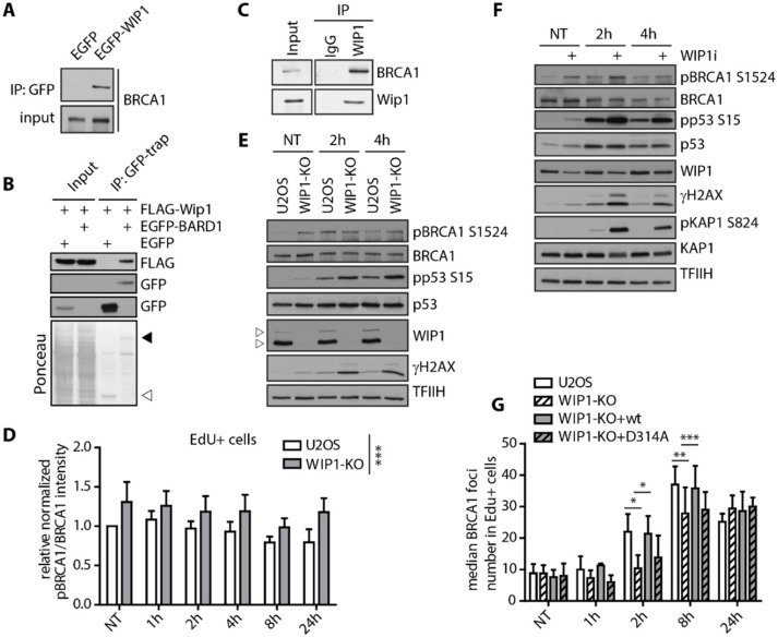
WIP1 interacts with BRCA1 and dephosphorylates S1524. (A) Co-immunoprecipitation of WIP1 and BRCA1. HEK293 cells were transfected with either empty GFP or GFP-WIP1, subjected to immunoprecipitation using GFP-Trap and analyzed by Western blotting with BRCA1 antibody. (B) Co-immunoprecipitation of WIP1 and BARD1. HEK293 cells were co-transfected with either empty GFP or GFP-BARD1 and Flag-WIP1, subjected to immunoprecipitation using GFP-Trap and analyzed by Western blotting with indicated antibodies. Ponceau staining with indicated positions of GFP (empty arrowhead) and GFP-BARD1 (full arrowhead) are shown. (C) Co-immunoprecipitation of endogenous WIP1 and BRCA1. U2OS cell lysates were incubated with 2 μg of a control antibody (IgG) or affinity-purified antibody against WIP1 for 2 h. Protein complexes were isolated by protein A/G resin and analyzed by immunoblotting. (D) Quantification of BRCA1 pS1524 signal intensity in replicating (EdU+) cells after irradiation. U2OS parental and WIP1 knockout cell lines were pulse-labeled with EdU for 30 min before irradiation. At indicated time-points, cells were pre-extracted, fixed and stained with pBRCA1 S1524 and BRCA1 antibodies. Click chemistry was used to visualize EdU. Median total intensity of BRCA1 pS1524 was normalized to total BRCA1 and is plotted +/− SD. Statistical significance evaluated by two-way ANOVA. (E) Western blot analysis of U2OS parental and U2OS-WIP1-KO cell lines after irradiation. Cells were irradiated and whole cell lysates were analyzed using Western blotting with indicated antibodies. Arrowheads indicate two isoforms of WIP1 present in U2OS. (F) Western blot analysis of MCF7 cells after irradiation with or without combined treatment with WIP1i. Cells were pretreated with WIP1 inhibitor for 30 min before irradiation and whole cell lysates were analyzed by Western blotting with indicated antibodies. (G) Quantification of BRCA1 foci in replicating (EdU+) cells after irradiation. Parental U2OS and U2OS-WIP1-KO cells and cell lines complemented with wild-type or phosphatase-dead (D314A) mutant of WIP1 were pulse-labeled with EdU for 30 min before irradiation. Cells were fixed after pre-extraction at indicated time-points and stained with BRCA1 antibody. Click chemistry was used to visualize EdU. Mean of median total intensity +/− SD is plotted. Statistical significance evaluated by two-tailed t-test.

