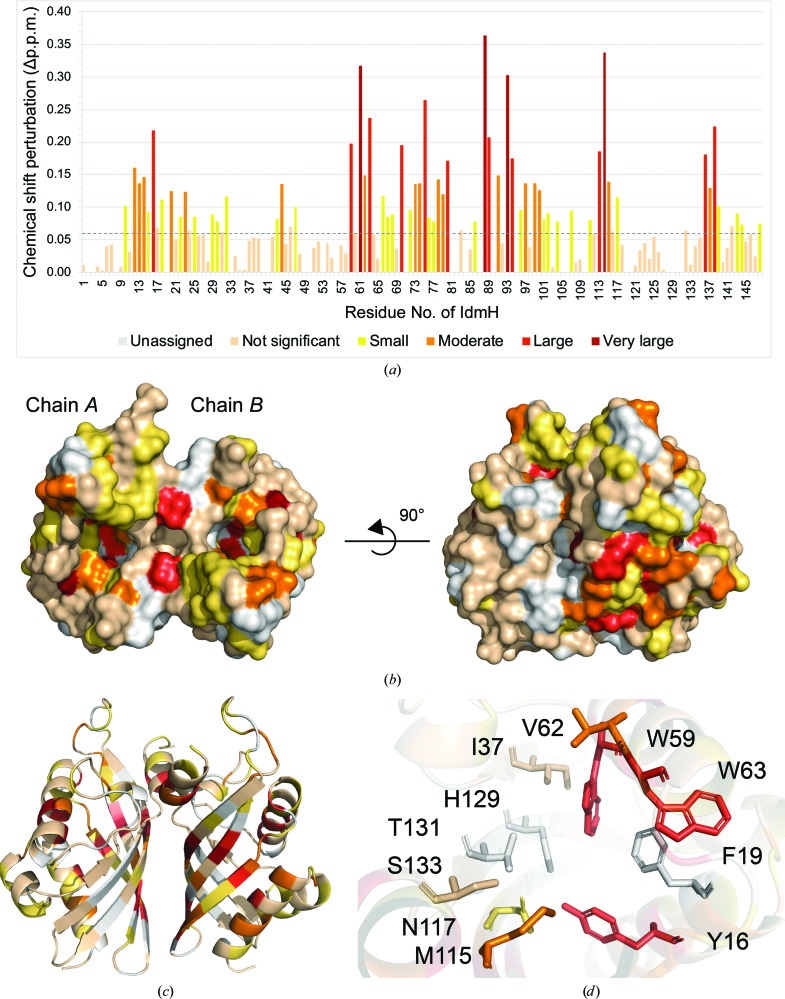
Figure 6.
Chemical shift perturbations (CSPs) mapped onto IdmH following the addition of indanomycin. (a) Histogram showing the geometrical distance moved by each peak assigned to a residue. Peaks are coloured from yellow to dark red as the value of the CSP increases. Chemical shifts which were smaller than the standard deviation of all shifts (0.072 p.p.m.) are coloured pale cream. Surface (b) and ribbon (c) representations of IdmH with peak shifts mapped using the same colour scheme as in (a). Residues which could not be assigned in the NMR spectrum are coloured white. (d) Active-site cavity of chain B with the proposed catalytic residues coloured according to the same colour scheme as in (b) and (c).

