
Figure 2.
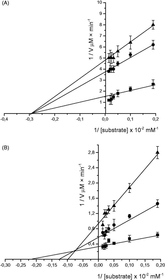
Lineweaver–Burk plot of glucosidase (A) and pancreatic lipase (B) activities. V is initial velocity and [S] is the concentration of substrate. The values were shown in absence (■) and presence of enzymatically modified chamomile infusion at 40 µM (●) and 60 µM (▲).The values are means of triplicate determinations, and the error bars indicate SD (n = 3).