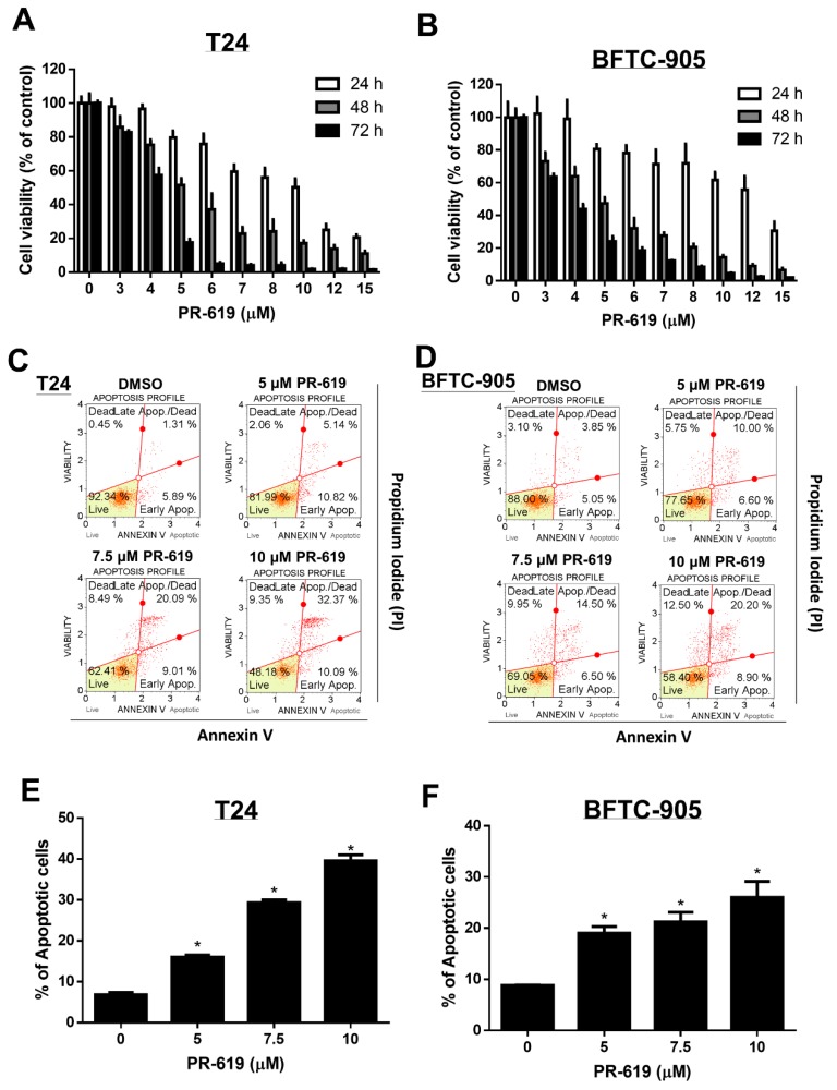
Figure 1.
PR-619 induced cytotoxicity and apoptosis in human urothelial carcinoma cells in a dose-dependent and time-dependent manner. (A) T24 and (B) BFTC-905 cells were treated with various concentrations of PR-619 (3–15 μM) for 24 h, 48 h, and 72 h, respectively. Cell viability was assessed using the 3-(4,5-dimethylthiazol-2-yl)-2,5-diphenyltetrazolium bromide (MTT) assay. (C) T24 and (D) BFTC-905 cells were exposed to PR-619 (5, 7.5, and 10 μM) or DMSO for 24 h. Apoptotic cells were analyzed through FACS flow cytometry with propidium iodide and annexin V-FITC staining. (E,F) show the quantitative analyses of apoptosis presented as the means ± SD; * p < 0.05 compared with controls. All results shown are representative of at least three independent experiments.

