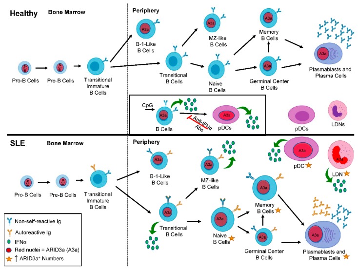
Figure 2.
ARID3a expression is enhanced in systemic lupus erythematosus (SLE) B cell subsets compared to healthy controls. A diagram depicts B lineage development in healthy (top panel) and SLE patients (bottom panel) from the bone marrow to the periphery. Red nuclei denote cell subsets that express ARID3a. The inset shows the induction of ARID3a and interferon alpha (IFNα; green circles), and the effect of those ARID3a-expressing B cells on healthy pDCs. In SLE patients, larger cells and gold stars indicate those cell subsets with increased numbers of ARID3a+ cells and associated IFNα production. Blue immunoglobulin (Ig) indicates non-self-reactive Ig, while gold Ig indicates the subsets of cells that can also produce autoreactive Ig. pDCs—plasmacytoid dendritic cells; LDN—low density neutrophils; A3a—ARID3a.

