Figure 6.
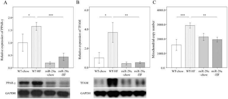
Overexpression of miR-29a modulates HFD-caused perturbation of mitochondrial biogenesis in the liver. Representative immunoblotting bands and densitometric results of (A) peroxisome proliferator-activated receptor γ (PPARγ) and (B) mitochondrial transcription factor A (TFAM), using GAPDH as the loading control. (C) mtDNA copy number per cell probed using qPCR, with TERT as the normalization control. Five to ten specimens were used for each group. Data are expressed as mean ± SE. * p < 0.05, ** p < 0.01, and *** p < 0.001 between the indicated groups. WT, wild type mice. HFD, high-fat diet. miR-29a, mice harboring overexpression of miR-29a.

