Figure 1.
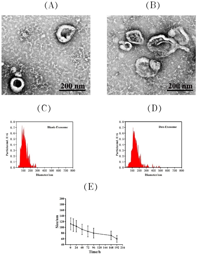
Characterization of exosomes: the representative TEM image of blank exosome (A) and exosome-doxorubicin (B). Size distributions of blank exosome (C) and exosome-doxorubicin (D) based on NTA measurements. The peak diameters were at 112.4 nm for free exosome and 152.7 nm for exosome-doxorubicin. (E) Particle size by nanoparticle tracking analysis for MSCs-derived exosomes during storage at −20 °C.

