
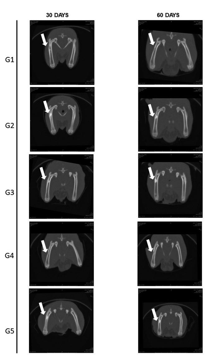
Figure 6. Representative images on coronal plane of the right femur of rats submitted to bone defect (white arrows) at 30 and 60 days after surgical procedure by CT scan.
Official websites use .gov
A
.gov website belongs to an official
government organization in the United States.
Secure .gov websites use HTTPS
A lock (
) or https:// means you've safely
connected to the .gov website. Share sensitive
information only on official, secure websites.