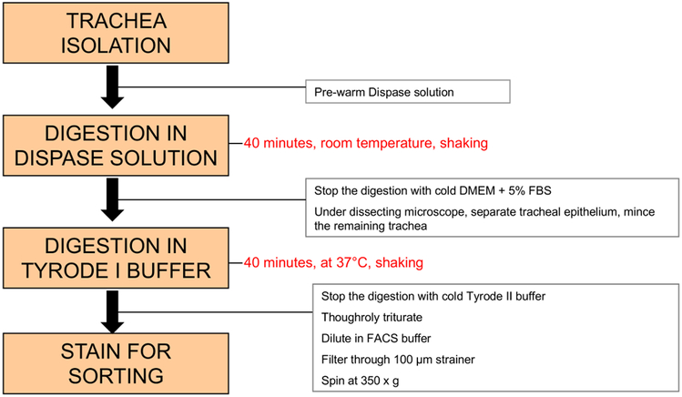
Figure 1: Schema of proposed steps for isolation of tracheal brush cells.
The protocol includes 2 major steps: after dissection, the whole trachea is incubated in a high-dose dispase solution to separate the epithelium; this is followed by digestion of the epithelial sheet with papain in calcium containing Tyrode buffer (Tyrode I) and antibody staining.

