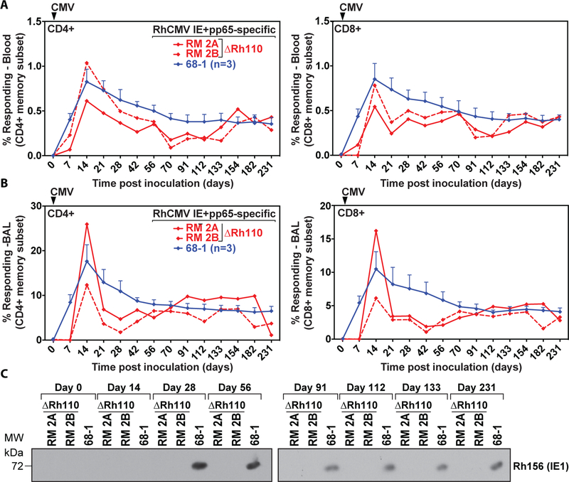
Figure 2: Rh110 (pp71)-deleted RhCMV retains T cell immunogenicity but is no longer shed in urine.
(A) Frequencies of RhCMV-specific CD4+ and CD8+ T cell responses in PBMCs were determined at the indicated time points in two RM inoculated with 107 PFU of ΔRh110 and three RMs given the same dose of 68–1 RhCMV. RhCMV IE1- and pp65a-specific T cells were determined by flow cytometric ICS after stimulation with mixes of consecutive, overlapping peptides comprising the RhCMV IE1 and pp65a proteins using intracellular expression of CD69 and either or both of TNF-α and IFN-γ to define Ag-specific T cells. Shown are response frequencies to IE1 and pp65a within the memory subset after background subtraction for each of the two ΔRh110-inoculated RMs (RM 2A and RM 2B) and the mean (+ SEM) of these response frequencies for the three RMs given 68–1 RhCMV. (B) Frequencies of RhCMV-specific T cell responses in bronchoalveolar lavages (BAL) were determined as in panel A in the same animals at the indicated time points. (C) Urine was isolated at the indicated days post-infection from RM 2A and RM 2B or one RM inoculated with 68–1. The presence of virus in co-cultures was determined by immunoblot for RhCMV IE1 (see Materials and Methods).

