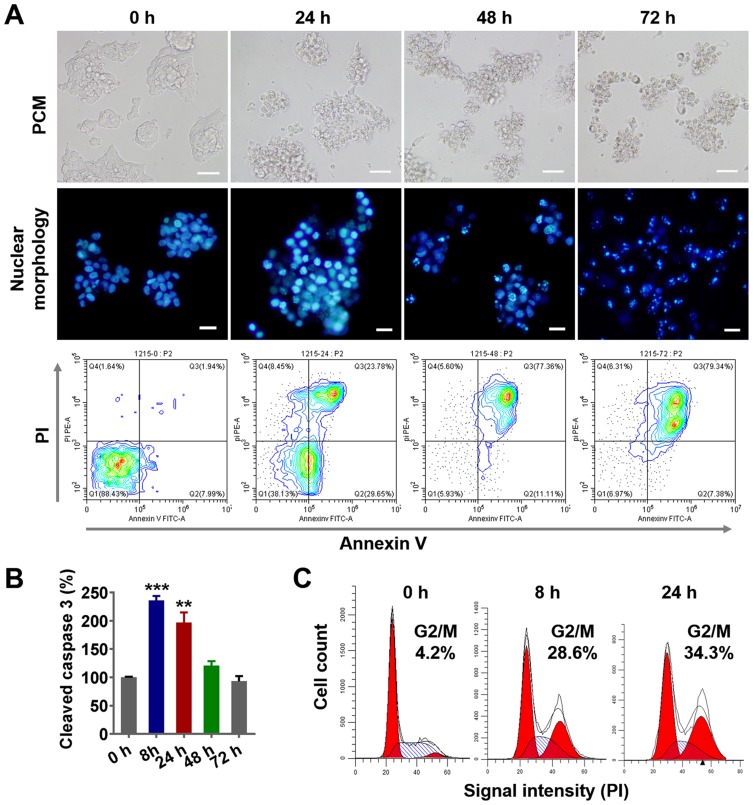
Figure 3.
PLGA-PTX-induced cell cycle arrest and tumor cell apoptosis.
Notes: Colorectal cancer cells (LS174T) were treated with PLGA-PTX over a time course. (A) Cell morphology was recorded by PCM, and cell nuclei were stained via Hoechst dye, and this was followed by fluorescence microscope imaging. Cell apoptosis was assayed by flow cytometry after dual staining with FITC-labeled Annexin V and PI. Scale bar, 50 μm. (B) Caspase 3 activation was analyzed by quantification of cleaved caspase 3 relative to a reference at 0 h. (C) Flow cytometry analysis revealed PLGA-PTX treatment induced accumulation of DNA from G0/G1 phase into G2/M phase over time following PI staining. **P<0.01; ***P<0.001.
Abbreviations: PLGA, poly lactic-co-glycolic acid; PTX, paclitaxel; PLGA-PTX, paclitaxel-loaded PLGA; PCM, phase contrast microscope; FITC, fluorescein; isothiocyanate; PI, propidium iodide.

