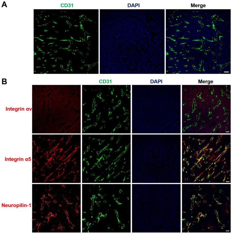
Figure 5.
Tumor vasculature distribution and expression of the iRGD receptor.
Notes: LS174T tumor frozen sections were prepared for immunofluorescence staining and imaging. (A) Blood vessel distributions were stained with anti-CD31 antibody (red). (B) The expression of iRGD receptors, including integrin αv, integrin α5, and neuropilin-1, were assayed by staining with anti-receptor antibodies (red). Tumor blood vessels (green) and nuclei (blue) were co-stained with anti-CD31 antibody and DAPI. Scale bar, 100 µm.
Abbreviations: CD31, cluster of differentiation 31; DAPI, 4,6-diamidino-2-phenylindole.

