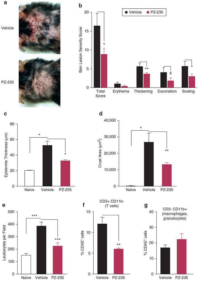
Figure 4. PZ-235 limits the severity of atopic dermatitis-like skin lesions in flaky tail mice treated with dust mite extract.
(a) Atopic dermatitis-like back skin lesions were induced in flaky tail mice (n = 6) by applying dust mite extract (40 μl, dose in mineral oil) twice weekly for 8 weeks with concurrent daily subcutaneous injections of PZ-235 (5 mg/kg) or vehicle. (b) Skin lesions were given a severity score based on erythema, skin thickness, scaling/lichenification, and scabbing. (c) Epidermal thickness and (d) skin crusting area were measured histologically in hematoxylin and eosin-stained sections. (e) Total dermal leukocytes in skin sections from flaky tail mice treated for 8 weeks with dust mites in a or naïve mice were identified by hematoxylin and eosin staining and morphology, and average numbers of leukocytes in 8 fields (original magnification × 20) per mouse quantified in a blinded manner. (f–g) CD45+ cells in digested fresh skin samples from dust mite-treated flaky mice in a were identified with CD45-APC-Ab and further stained for CD3-FITC-Ab and CD11b-APC-Cy7-Ab and quantified by FACS. Means ± standard error of the mean, n = 6 are shown. #P = 0.09, *P < 0.05, **P < 0.01, ***P < 0.001, ****P < 0.0001 versus vehicle by t test (b and f), or Dunnett test (c–e). Ab, antibody; APC, antigen-presenting cell.

