
Appendix Figure A1.
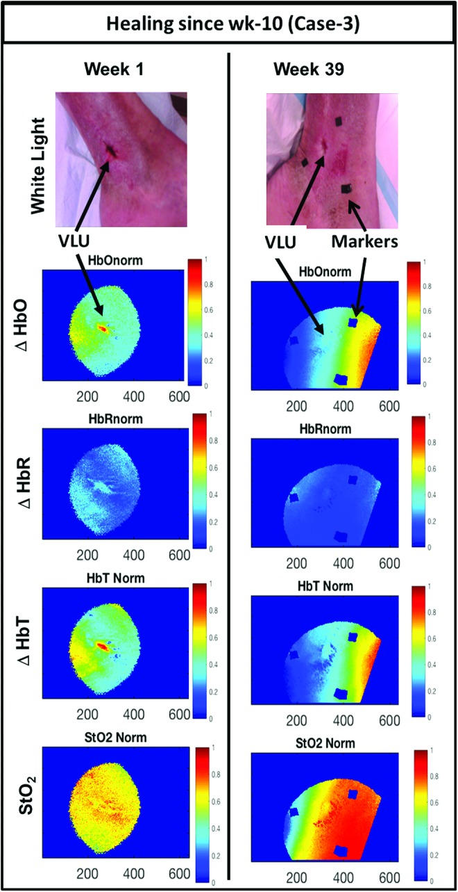
White light images (top row), normalized ΔHbO maps (second row), normalized ΔHbR maps (third row), normalized ΔHbT maps (fourth row), and normalized StO2 maps (fifth row) for healing VLU (case 3) at the first and last week of imaging. Color images are available online.