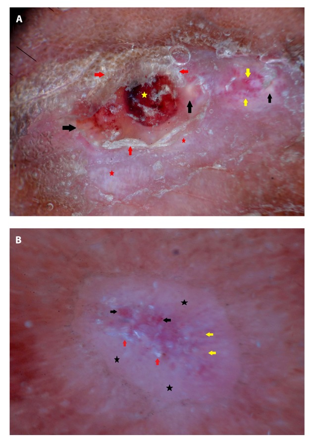
Figure 3.
Dermoscopy of eumycotic mycetoma; lesion 2: (A) Before treatment, yellow globules (black arrows), whitish structureless areas (red stars), telangiectasia and dotted vessels (yellow arrows), white superficial scales (red arrows), and blood spots (yellow star) are seen. (B) After treatment, reduction in yellow globules (yellow arrows), white superficial scales (red arrows), and dotted vessels (black arrows) are observed. Note the prominent whitish structureless areas (black stars) after treatment. [Copyright: ©2019 Ankad et al.]

