Figure 4.

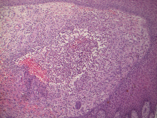
Histopathology of eumycotic mycetoma shows suppurative granuloma composed of neutrophils in the dermis (H&E, ×10). [Copyright: ©2019 Ankad et al.]

Histopathology of eumycotic mycetoma shows suppurative granuloma composed of neutrophils in the dermis (H&E, ×10). [Copyright: ©2019 Ankad et al.]