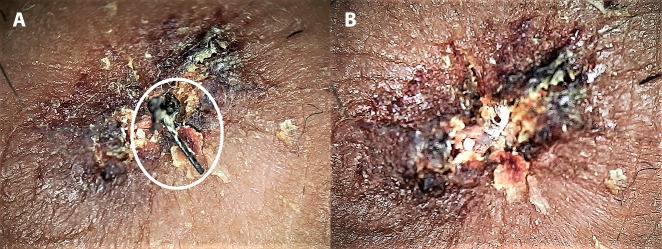
Figure 12.
Dermoscopy for identification and removal of retained sutures in a heavily crusted wound. (A) Before removal, the black suture, which was otherwise extremely difficult to delineate from the surrounding brownish black crust, is easily visible on dermoscopy (white circle). (B) After removal. (Escope video dermatoscope, Timpac Healthcare Pvt. Ltd., New Delhi, India; polarized, ×20.) [Copyright: ©2019 Sonthalia et al.]

