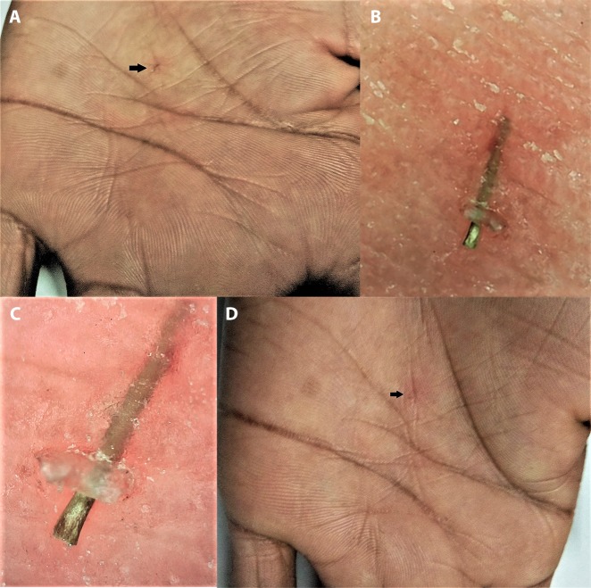
Figure 13.
Dermoscopy for identification and removal of intracutaneous foreign body. (A) Clinical image before removal—the tiny foreign body lodged intracutaneously (black arrow) with no visibility of protruding head or tail for removal. (B) Low magnification (×20) polarized dermoscopic image. (C) High magnification (×70) polarized dermoscopic image revealing the protruding head-end of the foreign body. (Escope video dermatoscope, Timpac Healthcare Pvt. Ltd., New Delhi, India; polarized, ×20.) (D) Clinical image after dermoscopically facilitated removal revealing a tiny linear subcorneal erosion (black arrow). [Copyright: ©2019 Sonthalia et al.]

