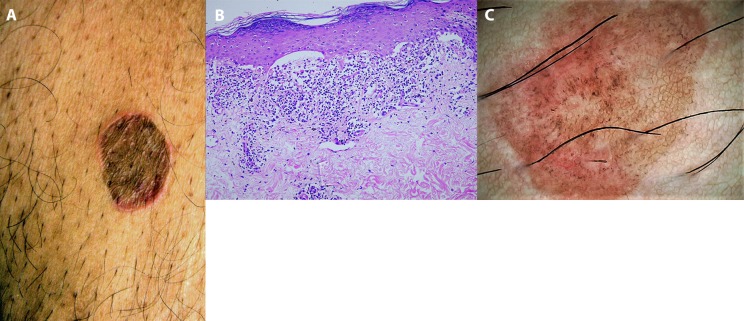
Figure 2.
Clinico-dermoscopic-pathological correlation. (A) Single, light brown, oval-shaped annular plaque over the lower anterior abdomen. (B) Histopathology suggestive of lichen planus, revealing mild hyperkeratosis, hypergranulosis with flattening of rete ridges, Max-Joseph space formation, dense bandlike lymphohistiocytic inflammation at the dermoepidermal junction, basal cell degeneration, and colloid bodies (H&E, ×400). (C) Dermoscopy revealed light brown pseudonetworks, overlapping pinkish areas, and multiple dark brown to blue-gray dots and globules. Wickham striae were not seen. Final diagnosis of early inflammatory lesion of lichen planus-like keratosis was confirmed. (Escope video dermatoscope, Timpac Healthcare Pvt. Ltd., New Delhi, India; polarized, ×20.) [Copyright: ©2019 Sonthalia et al.]

