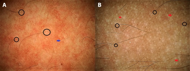
Figure 3.
Application of dermoscopy in suggesting disease activity in vitiligo. (A) A stable lesion of vitiligo fit for surgery displaying perifollicular depigmentation (black circles) and leukotrichia (blue arrow). (B) An unstable lesion displaying altered pigment network, “tapioca sago” appearance with multiple pearly-white dots (larger than eccrine openings), and micro-Köbner phenomenon (red arrows). (Escope video dermatoscope, Timpac Healthcare Pvt. Ltd., New Delhi, India; polarized, ×20.) [Copyright: ©2019 Sonthalia et al.]

