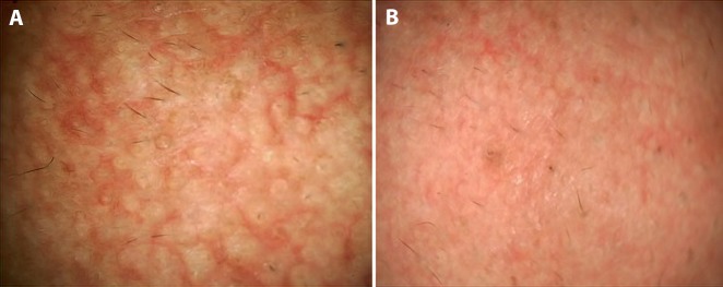
Figure 6.
Dermoscopy in early demonstration of treatment response in melasma. (A) Dermoscopic image from an untreated macule of melasma over the cheek of a woman displaying a diffuse nonspecific pattern of dark brown pigmentation with plentiful scattered dark brown globules and clods with perifollicular sparing and multiple prominent telangiectasias. (B) After 30 days of nightly local application of a 2% hydroquinone cream and daytime sunscreen and oral tranexamic acid (250 mg twice daily), dermoscopy from the same spot showing dramatic lightening of the background hue and erythema and reduction of pigmented structures and the telangiectasias. (Escope video dermatoscope, Timpac Healthcare Pvt. Ltd., New Delhi, India; polarized, ×20.) [Copyright: ©2019 Sonthalia et al.]

