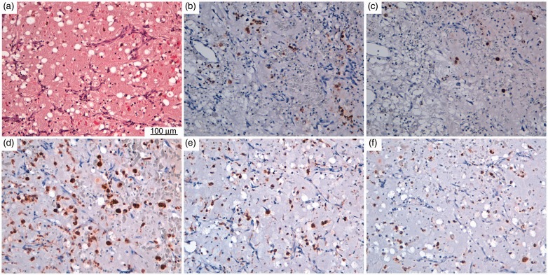
Figure 1.
(a) Histology and immunohistochemistry of mast cells and macrophages in myxoma samples. Histological slice stained with hematoxylin and eosin. Representative light microscopy images showing immunohistochemical detection (brown staining) of (b) c-kit (CD117), (c) tryptase, (d) CD68, (e) CD163 and (f) iNOS.

