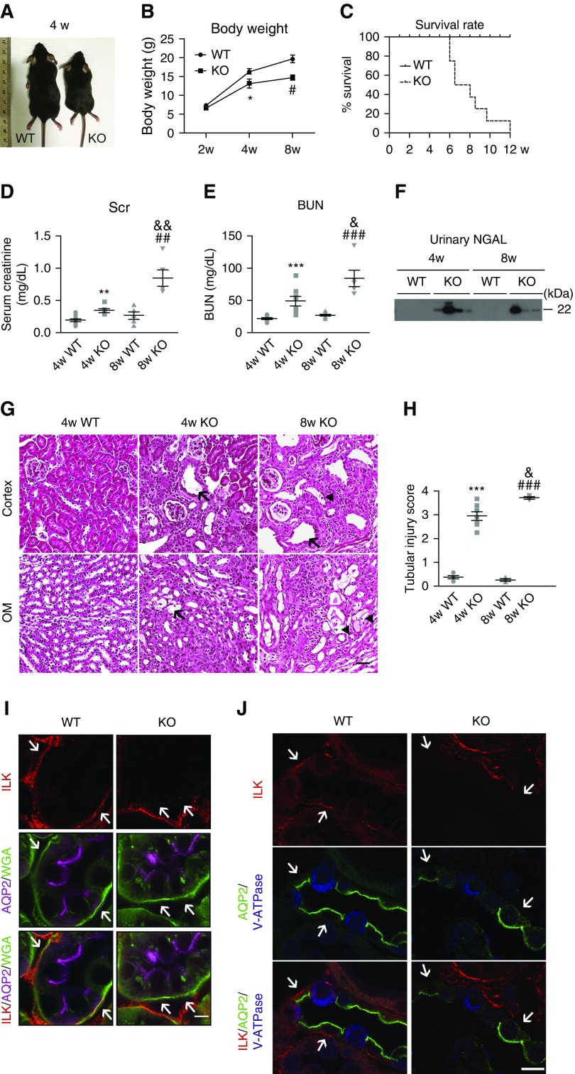
Figure 1.
Renal failure and kidney tubular injury are present in PC Ilk KO mice. (A) Representative images of wild-type (WT) and PC Ilk KO mice at 4 weeks old. (B) Body weight. *P<0.05 versus 4-week-old wild type; #P<0.05 versus 8-week-old wild-type mice. (C) Survival rate was shown by a Kaplan–Meier curve. n=8–10 mice per group. (D and E) Elevated serum creatinine (Scr) and BUN in PC Ilk KO mice. n=6–10. **P<0.01, ***P<0.001 versus 4-week-old wild-type mice; ##P<0.01, ###P<0.001 versus 8-week-old wild-type mice; &P<0.05, &&P<0.01 versus 4-week-old KO mice. (F) Increased urinary excretion of NGAL in PC Ilk KO mice as shown by immunoblotting. (G) Representative images of H&E staining of kidney cortex and outer medulla (OM) of wild-type and KO mice. Tubular dilation and microcysts were present in Ilk KO kidney (arrows). Detached and dead tubular cells were observed within tubular lumen (arrowheads). Scale bar, 50 μm. (H) Tubular injury score was calculated based on the percentage of damaged tubules. The degree of injury was graded blindly in ten randomly chosen fields as follows: 0, normal; 1, <10%; 2, 11%–25%; 3, 26%–75%; 4, >75%. ***P<0.001 versus 4-week-old wild-type mice; ###P<0.001 versus 8-week-old wild-type mice; &P<0.05 versus 4-week-old KO mice. n=4–7. Original magnification, 200×. (I) Immunofluorescence staining using an Airyscan confocal microscope confirmed deletion of ILK in PCs in KO kidney. ILK (red) was expressed on the basal membrane that was costained with wheat germ agglutinin (WGA)–FITC (green) in both PCs (stained purple with anti-AQP2 antibody) and ICs (negative for AQP2 staining) in wild-type kidney, whereas membrane expression of ILK (red) was significantly reduced or even absent in Ilk KO PCs. Arrows indicate ILK expression in WGA-positive basal membranes in wild-type and KO CDs, respectively. Scale bar, 5 μm. (J) Immunofluorescence staining revealed reduced ILK expression (red) in PCs in the KO kidney. PC-specific marker AQP2 stained green and IC-specific marker V-ATPase stained blue. ICs had low level of expression of ILK in general and there was no detectable change in ILK expression in KO kidney compared with the wild type. Arrows indicate ILK expression in basal membranes in wild-type and KO PCs, respectively. Scale bar, 10 μm. Data are presented as mean±SEM. Statistics were performed using the t test.

