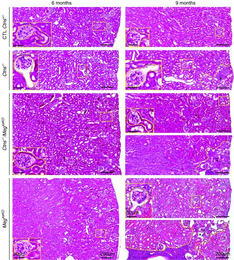
Figure 3.
Histology of cystinotic kidneys is preserved upon megalin ablation (whole-cortical views after staining with hematoxylin and eosin at 6 and 9 months). Broken yellow lines delineate inflammatory areas with parenchymal atrophy at 9 months (*). Representative GTJs are enlarged. In single Ctns−/− KO mice at 9 months, small foci of atrophy are always present but limited to the superficial cortex. For double KO and single MegksKO KO kidneys at 9 months, two examples are shown to illustrate either absence or presence of grossly remodeled areas that can span the entire cortex (see also Supplemental Figure 6B). CTL, control.

