Figure 4.
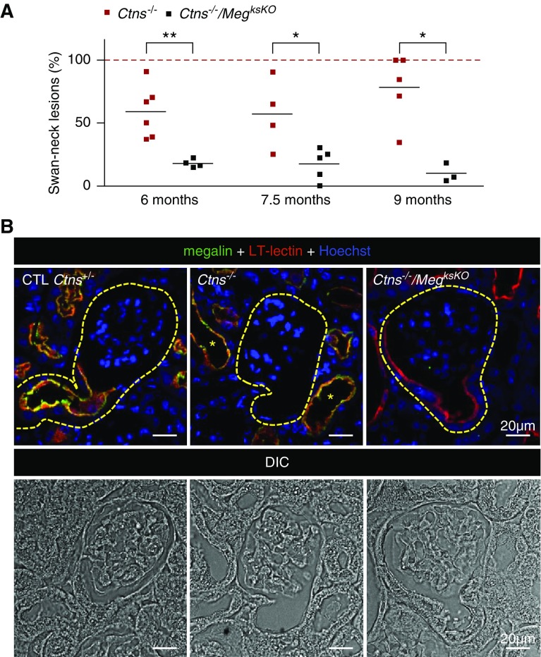
Megalin ablation in Ctns−/− kidneys prevents swan-neck lesions at GTJs. (A) Quantification of swan-neck lesions in Ctns−/− versus double KO mice from 6 to 9 months of age as percentage of all well defined GTJs over the entire sagittal kidney section, except at large inflammatory zones, as illustrated at Figure 3. *P<0.05, **P<0.01, nonparametric Mann–Whitney test). (B) Triple fluorescence confocal imaging with reference to differential interference contrast (DIC) imaging in Ctns−/− versus double KO mice at 6 months of age for L. tetragonolobus lectin (LT-lectin) labeling (red) and megalin (green) combined with nuclear Hoechst labeling (blue). Contours of GTJs are delineated by yellow broken lines. Notice in the central image that megalin is not detected at this representative Ctns−/− GTJ due to dedifferentiation/atrophy, but is preserved in more distal PTCs, together with LT-lectin labeling (yellow *). In contrast, the right image shows that megalin inactivation in Ctns−/− kidneys (no green signal) generally preserves PTC thickness and LT-lectin labeling at the GTJ. CTL, control.

