Figure 5.
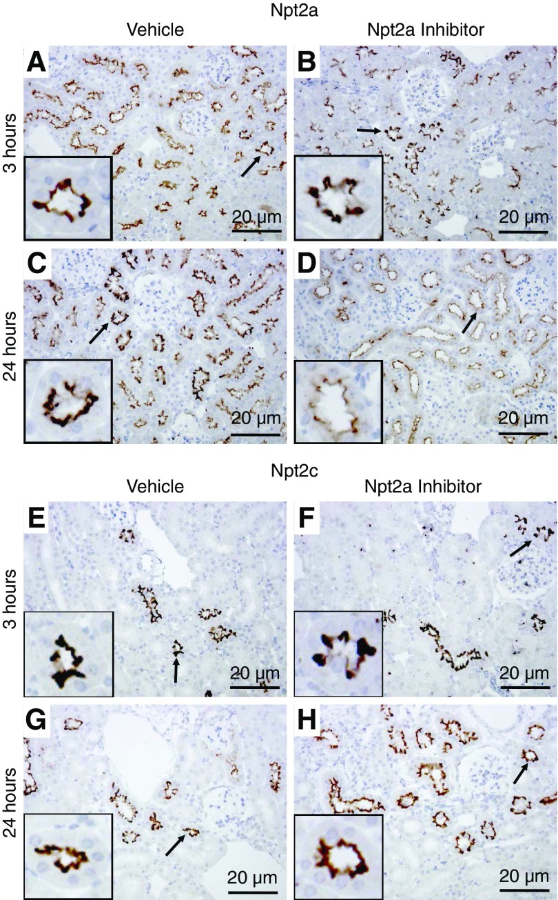
Qualitative immunohistochemical labeling identifies differences in Npt2a after 24 hours but not in Npt2c. Representative immunohistochemistry images after 3 and 24 hours from mice treated with vehicle or the Npt2a-I. (A) In vehicle-treated mice, Npt2a was detected throughout the proximal tubule starting in the early (S1) segment. Npt2a staining was predominantly localized to the apical microvilli. (B) Three hours after Npt2a-I treatment, Npt2a staining was not visibly different compared with vehicle treatment. (C and D) Twenty-four hours after treatment with the Npt2a-I, Npt2a staining was clearly weaker compared with vehicle. (E–H) Npt2c labeling was found throughout the proximal tubule in apical microvilli but staining intensity or distribution of Npt2c was not visibly different in vehicle- or Npt2a-I–treated mice after 3 or 24 hours. Arrows indicate regions magnified in the insets. n=5–6.

