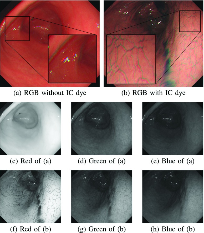
FIGURE 1.
Examples of endoscope images captured (a) without the IC dye and (b) with the IC dye. The color channel misalignment is observed in (a) and (b). The images (c) to (h) are six single-channel images extracted from (a) and (b). We can observe that the IC dye adds textures on the stomach surface, especially in the red channel (f).

