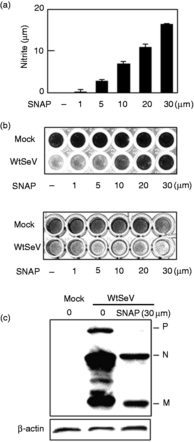
Figure 1.

Effect of NO on SeV multiplication. Following treatment with the indicated concentrations (a, b) or 30 μM (c) SNAP, one h before infection (b, upper panel, and c) or one h after infection (b, lower panel), Vero cells in a 96-well plate were mock-infected or infected with wtSeV at MOI 0.001 and incubated in 3 μg/ml trypsin. Culture media were collected 24 h postinfection and assayed for nitrite (a). Cells were also stained 72 h postinfection with 0.5% w/v amido black 10B dissolved in 20% ethanol and 10% acetic acid (b) or immunoblotted with rabbit serum against SeV (c). Viral proteins P, N, and M are marked in (c). SeV: Sendai virus; SNAP: S-nitroso-N-acetyl-DL-penicillamine; wt: wild type.
