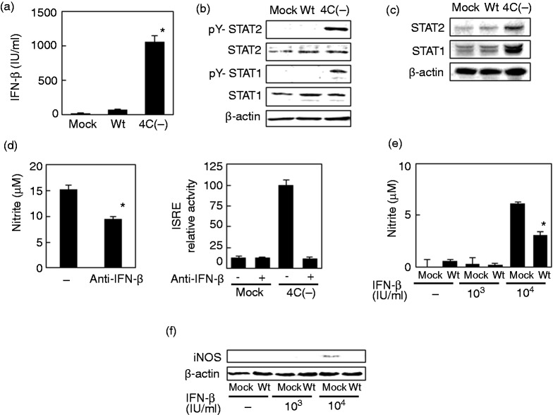
Figure 3.
Role of IFN-β in NO and iNOS production in RAW264.7 cells infected with SeV 4C(–). (a–c) Cells were mock-infected or infected with wt or SeV 4C(–) at MOI 5. Culture media were collected 24 h postinfection and assayed for IFN-β (a), whereas cells were harvested at five h (b) or 24 h postinfection (c) and immunoblotted with Abs to unphosphorylated and phosphorylated STAT1 and STAT2. (d) Cells were infected with SeV 4C(–) at MOI 5 and incubated for 24 h in the presence or absence of neutralizing Abs to IFN-β. Culture media were then assayed for nitrite. Cells were transfected with pRL-TK or pISRE-Luc, a firefly luciferase reporter plasmid driven by the ISRE promoter. At 24 h posttransfection, cells were mock-infected or infected with SeV 4C(–) at MOI 5, harvested after 8 h, and assayed by a dual-luciferase assay system to evaluate activation of the iNOS promoter. (e, f) Cells were mock-infected or infected with wtSeV at MOI 5. Cells were then mock-treated or treated with 103 or 104 IU/ml IFN-β for 20 h, beginning at 4 h postinfection. Culture media were then assayed for nitrite (e), whereas cells were immunoblotted with anti-iNOS (f). *P < 0.01 vs mock treatment with a-IFN-B antibody (d) or vs treatment with 104 IU/ml IFN-B after mock infection (e).iNOS: inducible NO synthase; ISRE: IFN-stimulated response element; SeV: Sendai virus; TK: thymidine kinase; wt: wild type.

