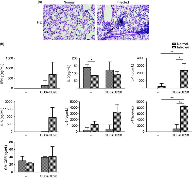
Figure 1.
The histopathological changes in the lung of infected C57BL/6 mice. (a) Sections of the lung of normal mice (left panels) and infected mice (right panels) were examined by H&E staining (×100). The multi-cellular granuloma could be observed in the infected group. (b) The levels of IFN-γ, IL-2, IL-4, IL-5, IL-6, IL-17, and GM-CSF were detected by CBA. The data are representative of six experiments, each with three or four replicates per group (*P < 0.05, **P < 0.01; the error bars indicate SD).

