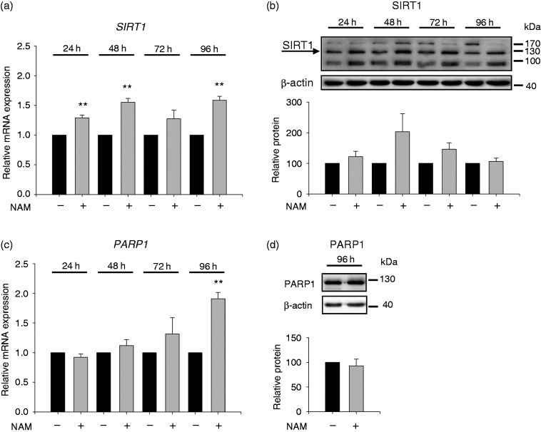
Figure 6.
SIRT1 and PARP1 expression after treatment with NAM. THP-1 cells (0.2 × 10⁶/ml) were treated with or without 8 mM NAM. After 24 h, 48 h, 72 h and 96 h mRNA values of (a) SIRT1 and (c) PARP1 were determined by real-time PCR. Data represent mRNA levels of NAM-treated cells relative to the untreated control at each time point analysed (-NAM = 1, black bar) ± SD (n = 3). (b) SIRT1 and (d) PARP1 protein were analysed by Western blot after 24 h, 48 h, 72 h, 96 h and 96 h, respectively. Densitometry data of proteins were normalised to the corresponding β-actin control. Data represent protein levels of NAM-treated cells relative to the untreated control at each time point analysed (-NAM = 100%, black bar) ± SD ((b) n = 3; (d) n = 4). Statistics were performed with the paired Student’s t-test calculated to the respective control.

