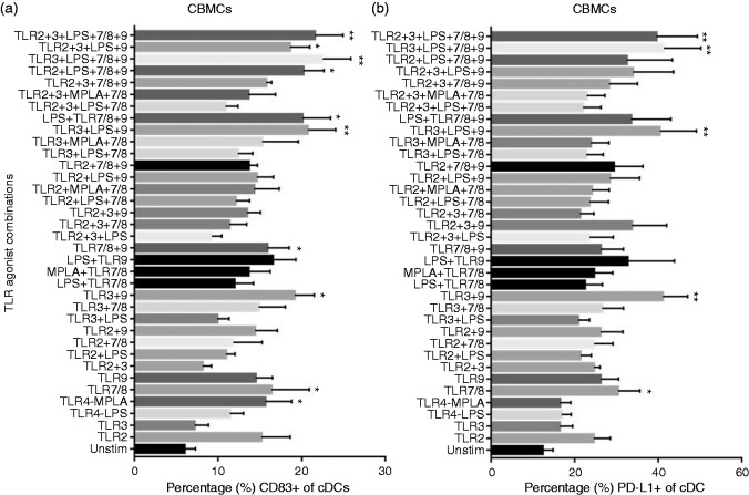
Figure 5.
Expression of costimulatory markers on neonatal classical dendritic cells in response to TLR stimulation. Cord blood (n = 8) mononuclear cells were stimulated as indicated. After 24 h, surface expression of (a) costimulatory marker CD83 and (b) inhibitory marker PD-L1 (CD274) were measured on neonatal classical dendritic cells by flow cytometry. Results are expressed as mean ± SE and analyzed using one-way ANOVA with Tukey’s post hoc correction for multiple comparisons. *P < 0.05, **P < 0.01, denotes significance compared to unstimulated control.

