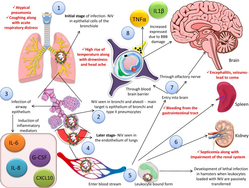
Figure 4.
Pathogenesis of NiV. 1. NiV can be seen in the epithelial cells of the bronchiole in the initial stage of infection. 2. NiV antigen can be detected in bronchi and alveoli. 3. Inflammatory mediators are activated as a result of infection to the airway epithelium. 4. Virus is disseminated to the endothelial cells of the lungs in the later stage of the disease. 5, 6. Virus enter the blood stream followed by dissemination, either freely or in host leukocyte bound form, reach brain, spleen and kidneys. 7. Two pathways are involved in the process of viral entry into the central nervous system (CNS), via hematogenous route and anterogradely via olfactory nerve nerves. 8. The blood brain barrier (BBB) is disrupted and IL-1β along with tumor necrosis factor (TNF)-α are expressed due to infection of the CNS by the virus which ultimately leads to development of neurological signs. Red font shows the symptoms in human.

