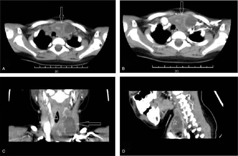
Figure 1.

A, CT scan showing a heterogeneous cystic and solid mass (white arrow) within the left thyroid lobe and clear margins that contained calcifications. B, Intravenous contrast-enhanced axial reveals no evident enhancement,(white arrow). C, There are distortion and compression of the airway (white triangle). D, Coronal views. CT = computed tomography.
