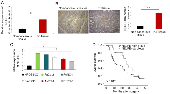
Figure 1.
Increased NELFE expression in PC tissues and cells. (A) The expression of NELFE in samples was analyzed by reverse transcription-quantitative PCR. (B) Analysis of the expression of NELFE in samples using an immunohistochemistry assay. The positive expression of NELFE was brown and yellow. The nuclei of cells were blue. (C) NELFE expression levels in various human PC cell lines and HPDE6-C7 cells. (D) Comparison of Overall survival between PC patients with high and low NELFE expression levels (n=120). The 120 pair of PC tissues and adjacent non-tumor clinical samples were removed from patients with PCs. *P<0.05 vs. HPDE6-C7; **P<0.01 vs. non-cancerous tissues or NELFE-low group. NELFE, negative elongation factor E; PC, pancreatic cancer.

