
Figure 1.
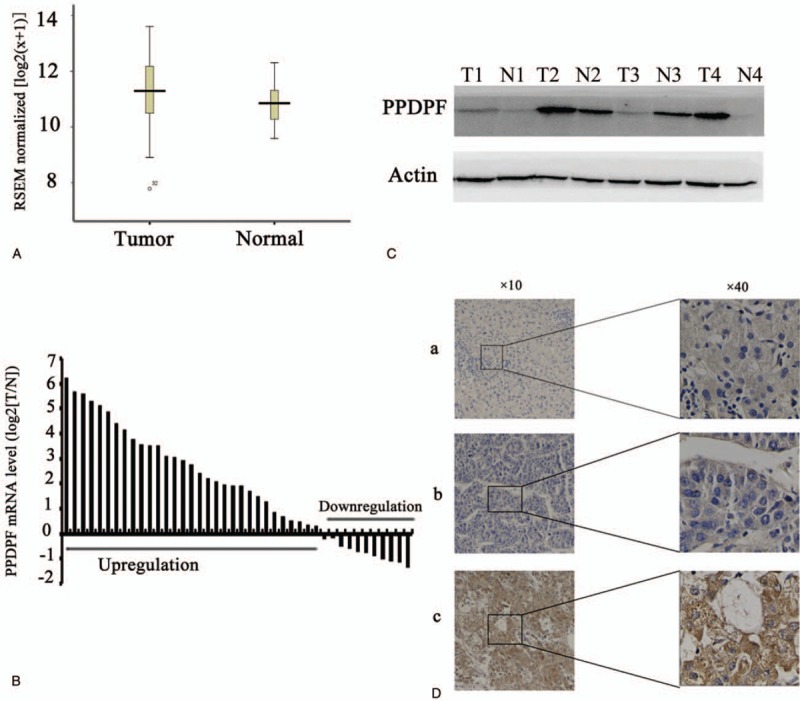
PPDPF expression is upregulated in HCC. (A) Analysis from the TCGA database (http://xena.ucsc.edu/) shows that mRNA expression levels of PPDPF is significantly higher in HCC compared with normal liver tissues. (B) Quantitative real-time PCR results of the relative expression level of PPDPF in 42 pairs of HCC and normal liver tissue samples. All of the reactions were performed in triplicate and results represent the mean ± SD (P < .05). The expression of PPDPF is normalized to beta-actin. (C) Expression of PPDPF protein in 4 randomly selected paired HCC samples was analyzed using western blot. N, normal; T, primary HCC. (D) Immunohistochemistry (IHC) results of PPDPF in human HCC specimens and adjacent normal tissues. a: expression of PPDPF in the paired adjacent tissues; b: low expression of PPDPF in HCC tissue; c: High PPDPF expression in HCC tissue. HCC = hepatocellular carcinoma, PCR = polymerase chain reaction, PPDPF = pancreatic progenitor cell differentiation and proliferation factor.