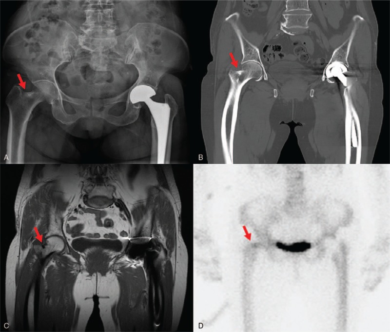
Figure 1.

Preoperative images showing incomplete fracture of lateral cortex of femoral neck (red arrows). A, X-ray. B, CT scan (coronal view). C, T1-weighted MR image (coronal view). D, bone scan image showing small focal increased bone uptake at the fracture site.
